
|
|
Papillary carcinoma - Case 57.
|
|


Clinical data: a 23-year-old woman was evaluated because of diffuse non-specific complaints. The ultrasonography disclosed a nodular goiter.
Palpation: a moderately firm nodule in the isthmic part of the left lobe.
Functional state: euthyroidism (TSH 2.23 mIU/L, FT4 15.0 pM/L).
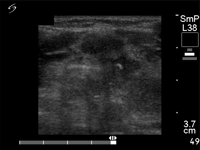
Ultrasonography: both thyroids were minimally hypoechogenic. There was a hypoechogenic nodule in the isthmic part of the left lobe. The nodule showed increased blood flow and had irregular borders.
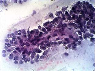
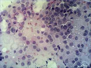
Cytological picture: no colloid in the background. Thyrocytes in papillary clusters, forming microfollicles and dissociated. They have abundant cytoplasm, and exhibit nuclear inclusion. A relatively great number of lymphocytes are present on the smear.
Cytological diagnosis: papillary cancer .
We performed anti-TPO determination which resulted in almost normal level 46 U/L.
Histopathological diagnosis: papillary cancer, Hashimoto's thyroiditis.
.