
|
|
Chronic lymphocytic thyroiditis - Case 73.
|
|


First examination (1st row of images):
Clinical presentation: a 63-year-old woman came to a yearly follow-up examination because of a known thyroid nodule and a hypothyroidism replaced with daily 75 microgram levo-tiroxine. She had neck discomfort She had neck discomfort while turning her head to the right.
Palpation : there was a nodule in the left lobe.
Hormonal investigation: indicated euthyroidism with TSH-level 1.01 mIU/L.
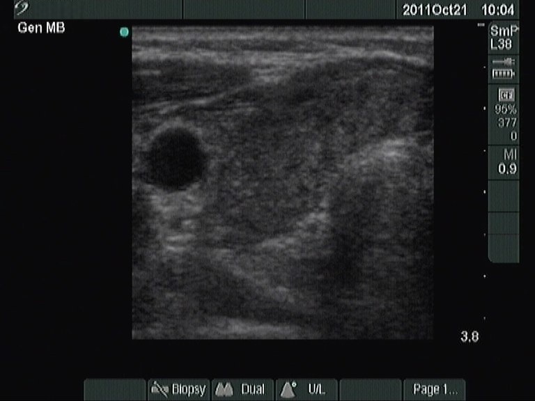
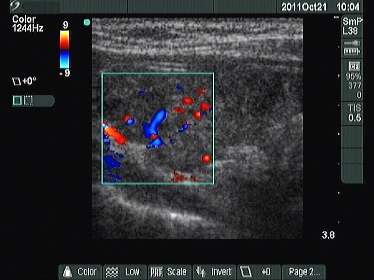
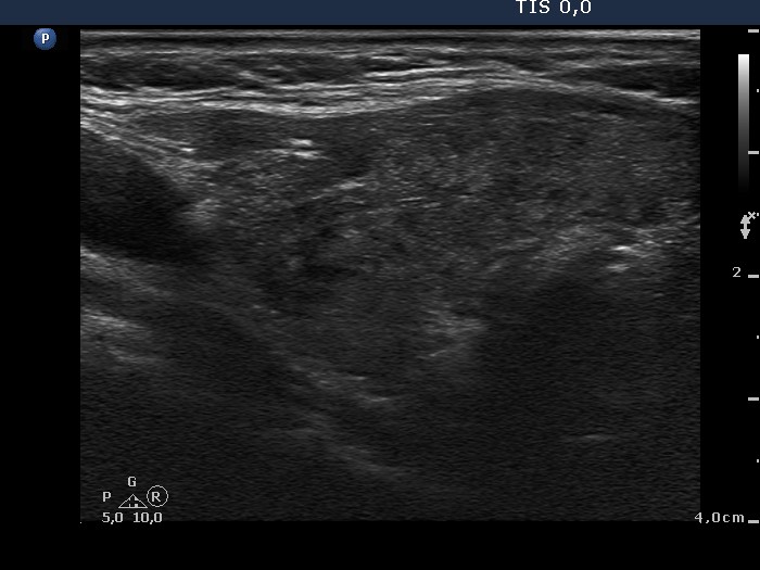
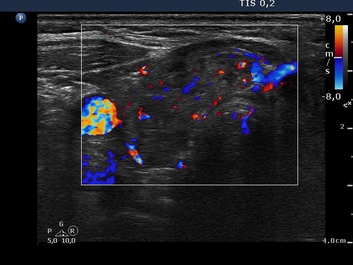
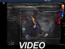
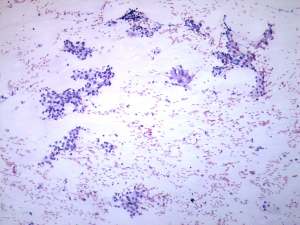
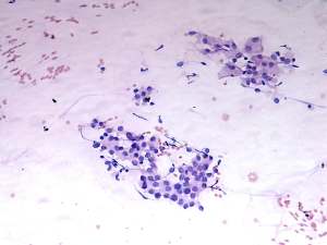
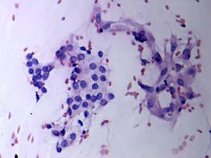
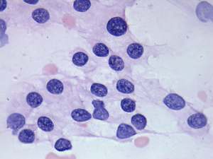
Ultrasonography: revealed hypoechogenic inhomogeneous thyroids. There was a hyperechogenic nodule in the left lobe. The nodule presented a halo sign and perinodular blood flow. It increased in size, therefore we performed aspiration cytology.Cytology: benign lesion.
X-ray examination excluded tracheal compression and neck rib. We offered rheumatological examination.
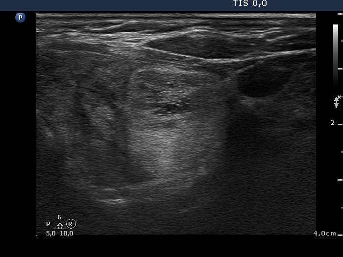
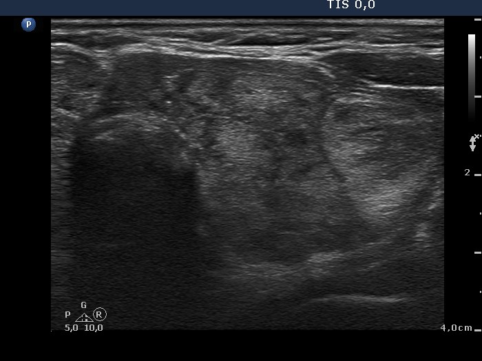
Follow-up examination 2 years later (2nd and 3rd rows of images):
Clinical presentation: the complaints of the patient worsened and requested a repeat examination.
Palpation: there was a nodule in the left lobe.
Hormonal investigation: indicated euthyroidism on daily 87.5 microgram levothyroxine (TSH-level 1.88 mIU/L).
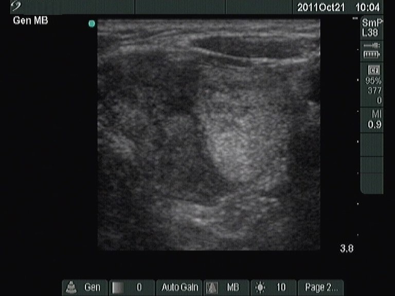
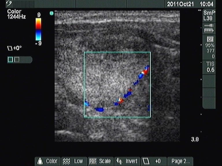
Ultrasonography: was unchanged except for the increase of the left lobe by 38% in volume.Cytology: benign lesion.
The patient was operated on because of compression signs.
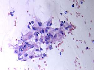
Histopathology: Hashimoto's thyroiditis and multiple hyperplastic nodules in the left lobe.
Comments.
-
It is worth comparing the numerous small echonormal lesions and the large nodule in the left thyroid. The former are part of the so-called pseudonodular form of Hashimoto's thyroiditis, while the true nodule was much larger. Pseudonodules are usually in the range of 5 to 15 mm in maximal diameter.
-
This patient had a rare compression sign. We suppose that the nodule comprised a nerve while the patient turned her head to the right. This complaint was resolved after the surgery.
-
Most solitary nodules which are greater than 2 cm in maximal diameter, display a halo sign and perinodular blood flow are proved to be follicular tumor. This case belongs to the relatively rare exceptions.