Benign nodular hyperplasia - Case 56. (ultrasonographic picture 5)
|
|

|
|
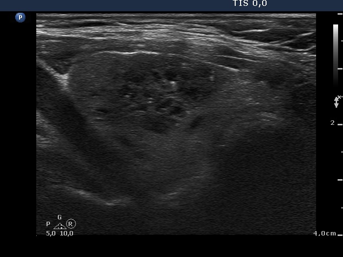
Lower 1/3 of the right lobe, longitudinal scan. Note comet-tail artifacts within the cystic lesion.

|
|
|
|
Lower 1/3 of the right lobe, longitudinal scan. Note comet-tail artifacts within the cystic lesion.