Benign nodular hyperplasia - Case 56. (ultrasonographic picture 6)
|
|

|
|
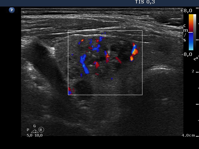
Middle part of the right lobe, horizontal scan, color Doppler mode. Type 3 vascular pattern.

|
|
|
|
Middle part of the right lobe, horizontal scan, color Doppler mode. Type 3 vascular pattern.