
|
|
Teamwork - case 643
|
|


Clinical data. A 35-year-old woman discovered a lump in the right thyroid lobe several months ago.
Palpation: There was a not firm nodule in the right lobe.
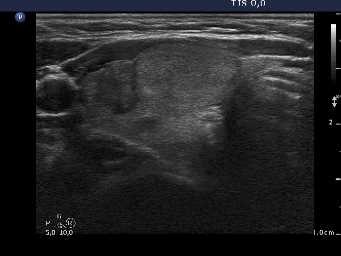
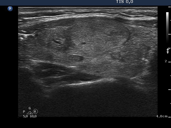
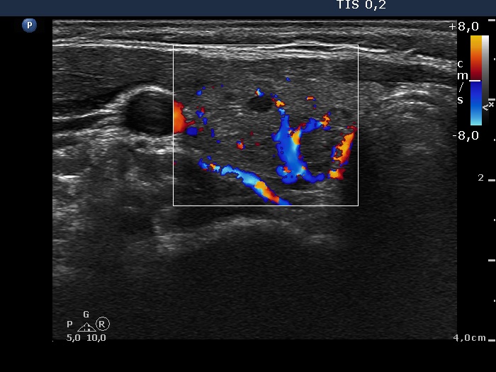
Ultrasonography. The thyroid was echonormal. There were multiple moderately hypoechogenic lesions in the right lobe. The palpable nodule was echonormal and presented halo sign and perinodular blood flow.
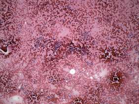
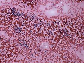
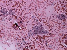
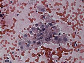
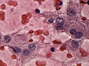
Cytology was performed from one of the moderately hypoechogenic lesions. It was located next lateral to the echonormal lesion. There were two cell populations on the smear: beside regular thyrocytes, follicular cells presenting oxyphilic metaplasia were also found on the smear.
Functional state: euthyroidism (TSH 1.30 mIU/L). Anti-TPO was not in the normal range (9 U/mL).
Combined cytological-clinical-sonographic diagnosis: benign lesion presenting oxyphilic metaplasia.
Because of cosmetic reasons, the patient wished to be operated.
A right lobectomy was performed. Histopathology disclosed benign hyperplastic nodules and chronic lymphocytic thyroiditis.
Comments.
-
The possibility of an oxyphilic tumor could not be fully excluded solely on cytology.
-
Taking the ultrasound presentation into account, the risk of oxyphilic tumor was even lower because the ultrasound pattern corresponds either to hyperplastic nodules or to an autoimmune thyroiditis.
-
Although the possibility of an autoimmune thyroiditis could not be excluded on the ultrasound presentation, the normal anti-TPO level and the lack of lymphocytes on the smear stood against Hashimoto's thyroiditis.