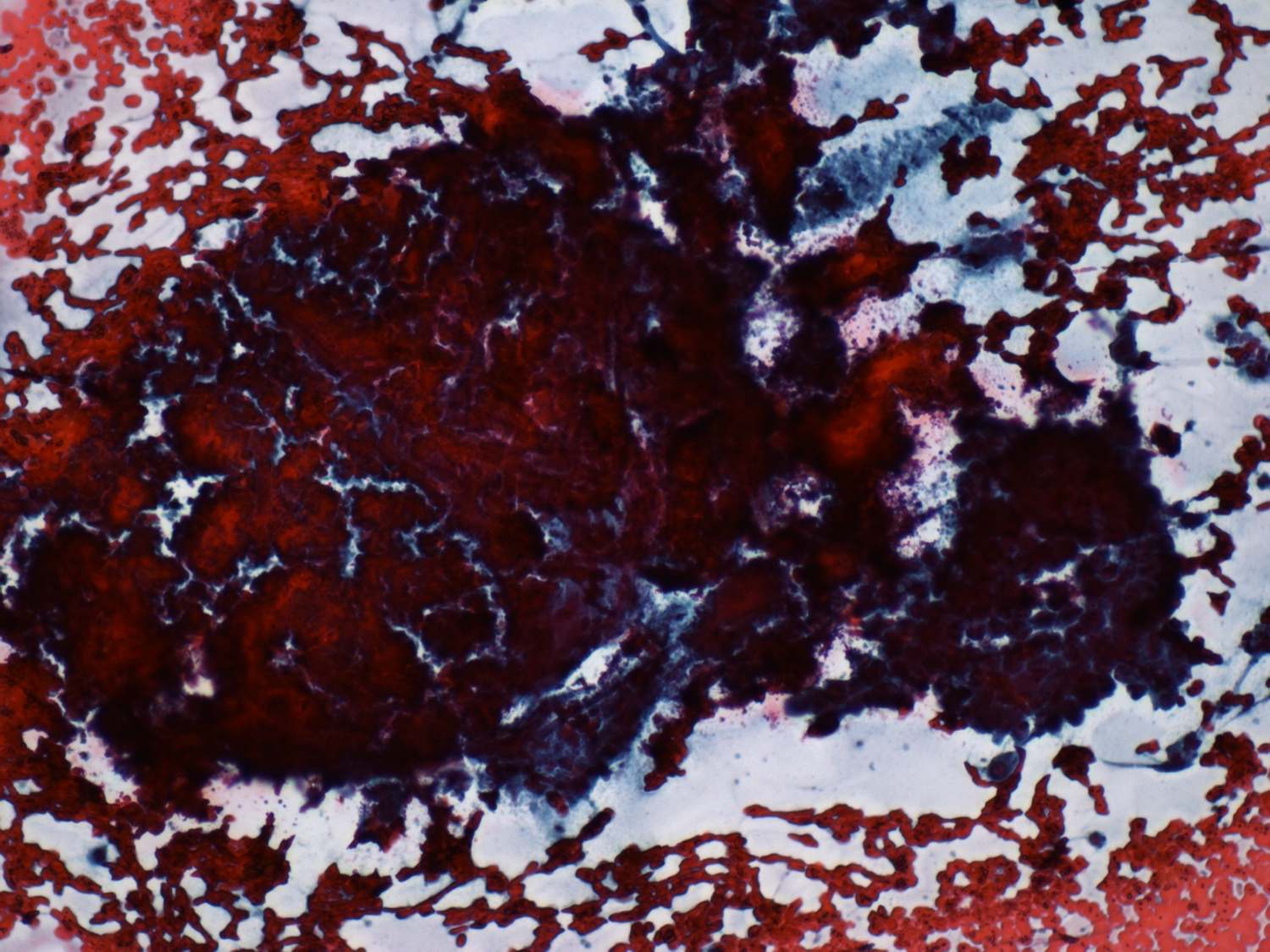
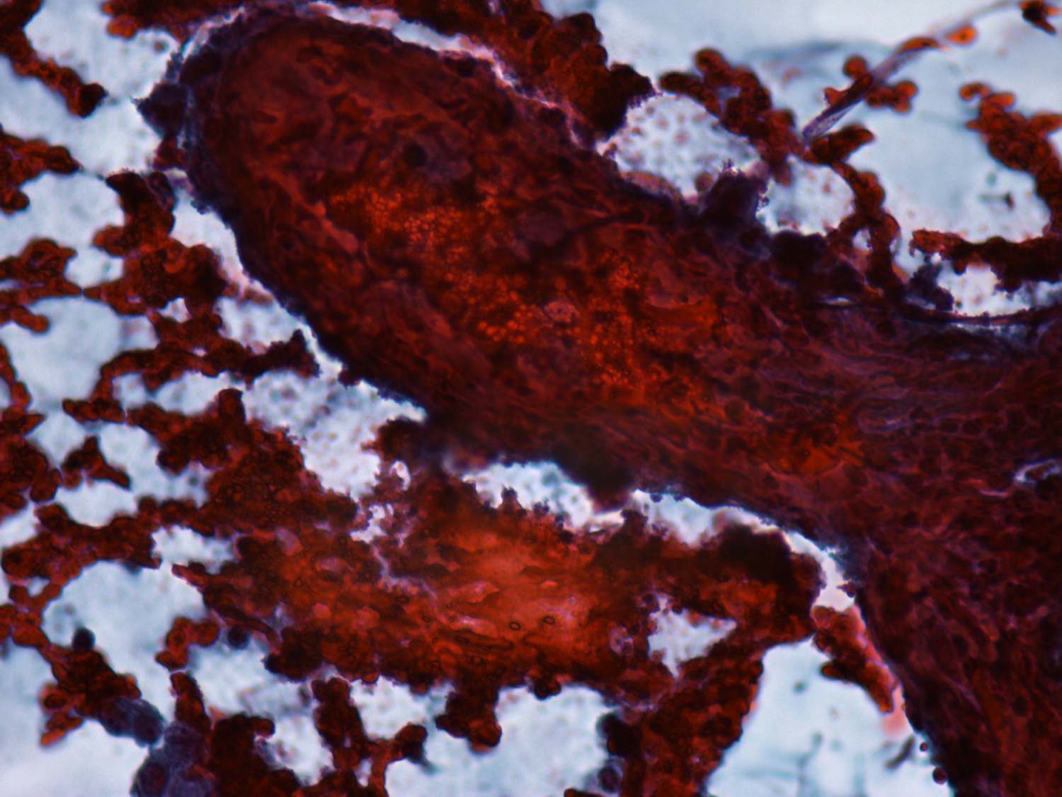
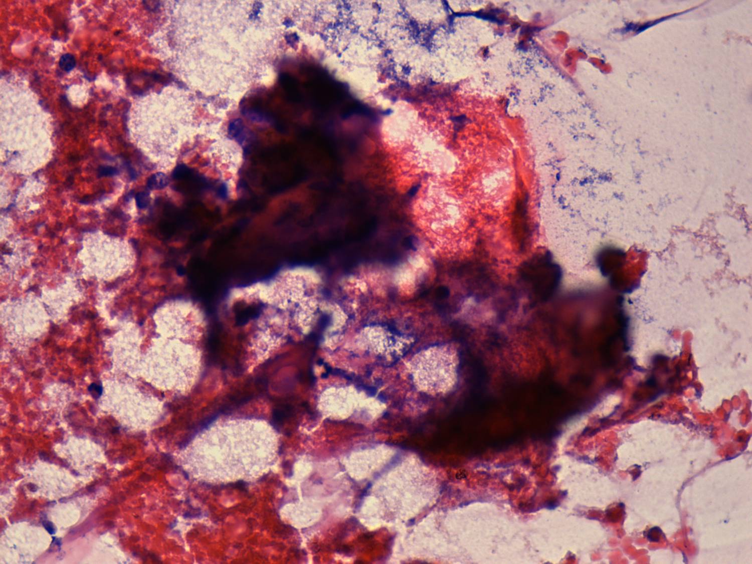
Psammoma body is considered to be the most characteristic and
specific
cytological feature of a papillary cancer. It corresponds to a
calcified
papillary structure and has a concentric appearance. It is best to
observe by finely varying the focus during microscopic analysis: we can
see several concentric figures in different depth.
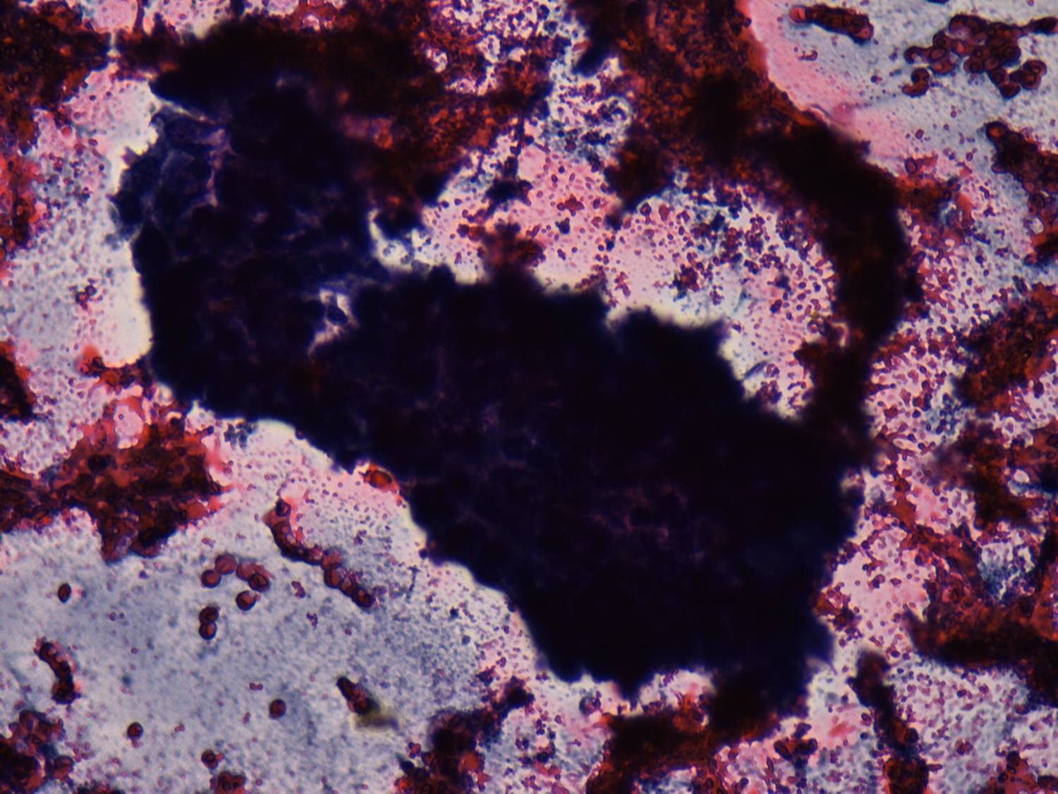
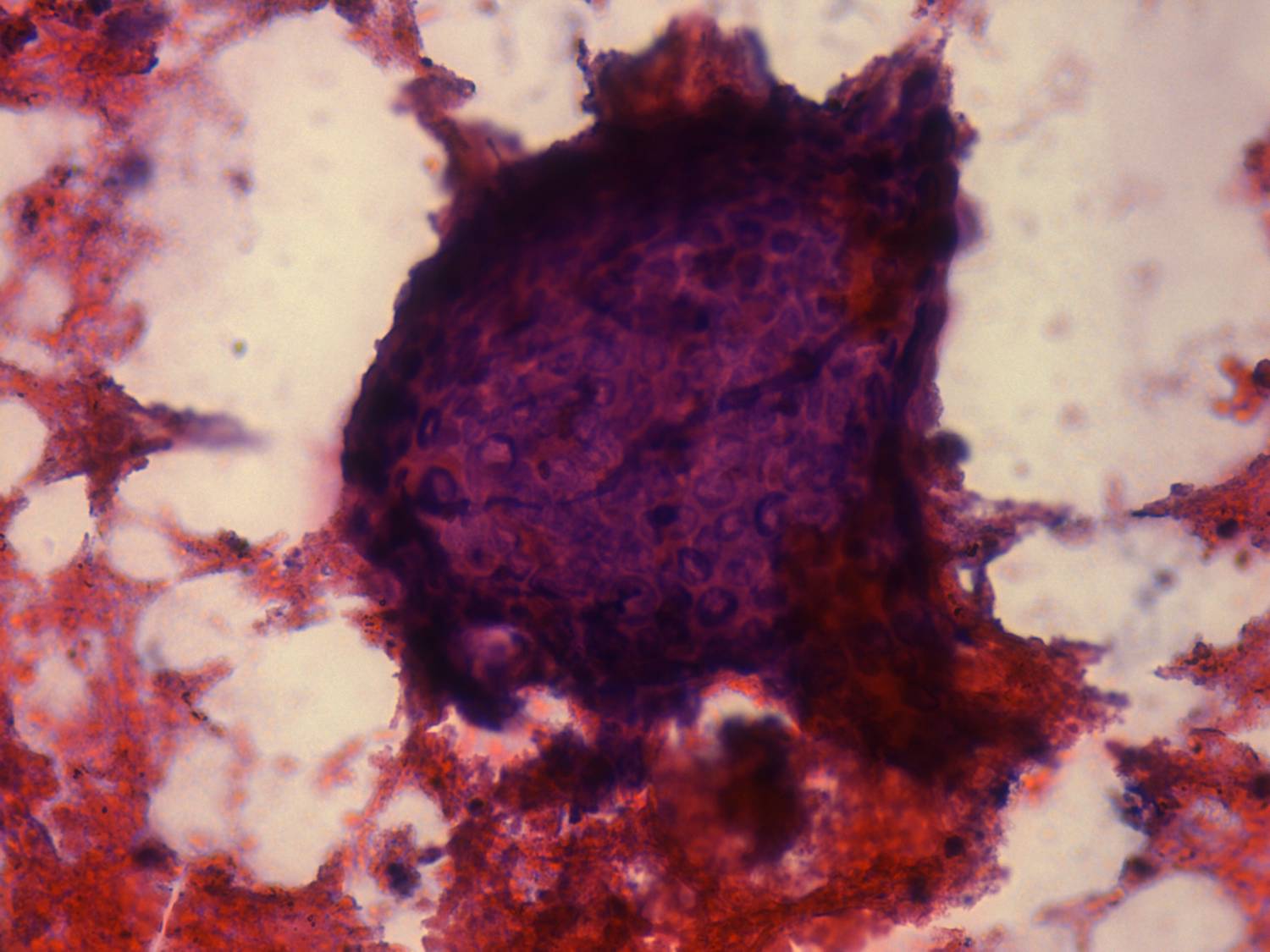
The occurrence of psammoma body is related to the size of the needle
used during aspiration, because psammoma body is a relatively large
structure. We prefer to use thin needles (23 G and 25 G) in order to
avoid bloody material, therefore we have found psammoma bodies in less
than 2% of papillary cancers.