
QUICK TOUR - Case studies - sample case 3 of 6:Papillary carcinoma - Case 11. (ultrasonographic picture 6)
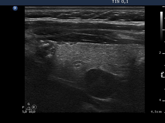 Left lobe, longitudinal scan. The papillary carcinoma is in the upper pole (left in the image) while the cystadenoma is in the dorsal part of the lobe. |
