
QUICK TOUR - Case studies - sample case 3 of 6:Papillary carcinoma - Case 11. (ultrasonographic picture 7)
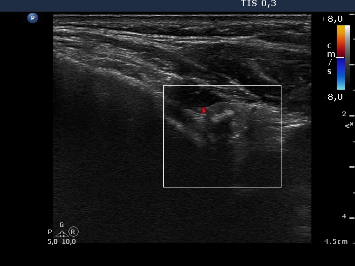 Upper pole of the left lobe, horizontal scan, color Doppler mode. A type 1 vascular pattern. |
QUICK TOUR - Case studies - sample case 3 of 6:Papillary carcinoma - Case 11. (ultrasonographic picture 7)
Upper pole of the left lobe, horizontal scan, color Doppler mode. A type 1 vascular pattern. |
