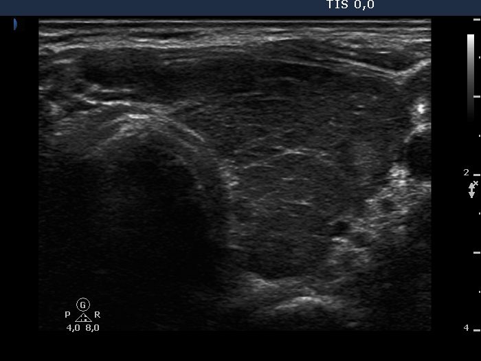
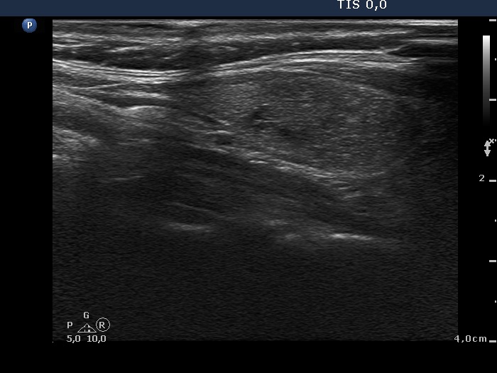
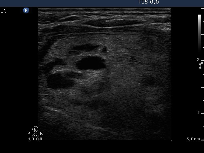
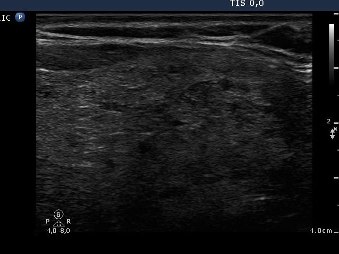
Benign Hashimoto's thyroiditis (cytological diagnosis) - case 679 |
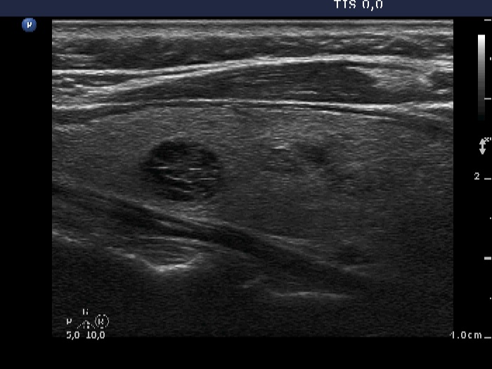
Benign Graves' disease (clinical diagnosis) - case 543
|
 |
 |
 |
 |
First, we demonstrate two cases of autoimmune thyroid diseases with fibrotic changes. Both present pale and bright hyperechogenic granules and lines corresponding to normal and excessive connective tissue. Neither of the discrete areas are nodules in a pathological sense.
|
| |
|
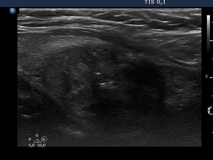
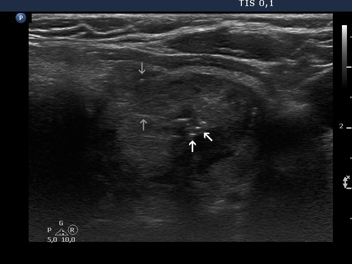
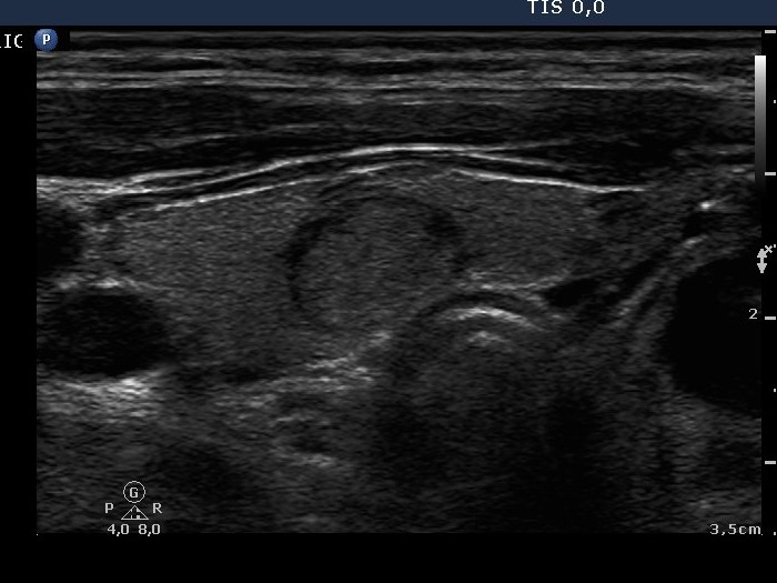
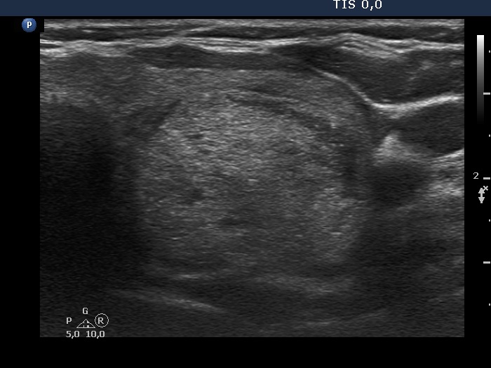
Benign colloid goiter (cytological diagnosis) |
 |
 |
 |
 |
The nodule has both pale and bright hyperechogenic lines and granules. Arrowheads point to figures representing the normal architecture of the thyroid while arrows do to thickened connective tissue.
|
| |
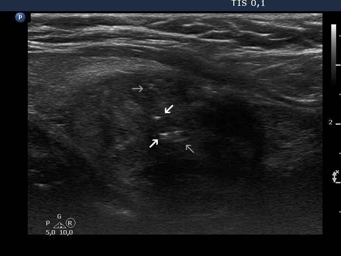
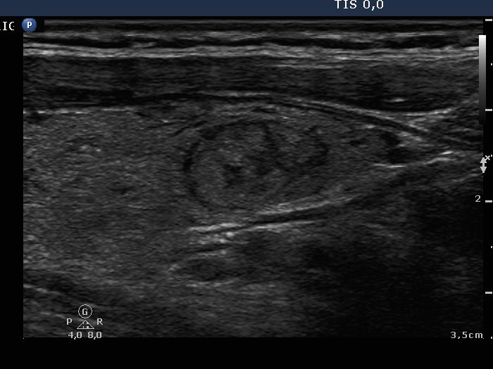
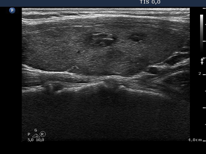
Benign colloid goiter (cytological diagnosis) - case 1429 |
 |
 |
The nodule has pale hyperechogenic granules and lines corresponding to the normal architecture of the thyroid consisted of connective tissue.
|
| |
|
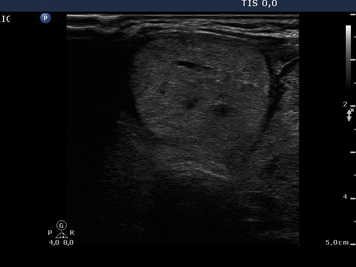
Intact right lobe without any pathological finding and benign hyperplastic nodules in the left lobe (histological diagnosis) - case 803 |
Right lobe |
 |
 |
Left lobe
|
 |
 |
Both the pseudonodule in the right lobe (upper images) and the benign nodule in the left lobe (lower images) have numerous pale granules and lines which correspond to the normal architecture of the thyroid and correspond to connective tissue. The finding of a few more bright granules and lines are the ultrasound presentation of thickened connective tissue.
|
| |
|
Benign cystic-colloid goiter (cytological diagnosis) - case 1473
|
 |
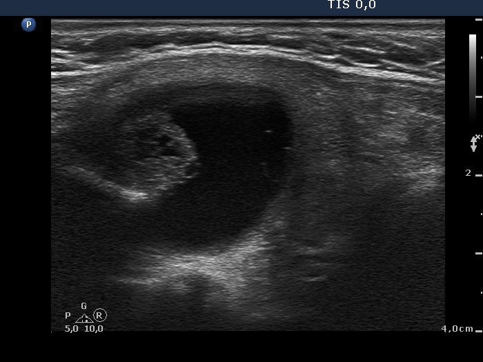 |
The solid part has pale and bright granules and lines which correspond to connective tissue or back wall figures.
|
| |
|
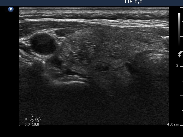
Benign lesions (cytological diagnosis) - case 1644 |
Right lobe |
Left lobe |
 |
 |
 |
 |
Back wall cystic figures are presented in the left images, while the origin of the hyperechogenic figures in the right image is in part ambiguous: several of them are located in the back wall of tiny cystic areas, others seem not to be related to cystic fluid. The latter are therefore presentations of connective tissue.
|
| |
|
| |
|
Benign lesions (cytological diagnosis) - case 808
|
Right lobe |
Left lobe |
 |
 |
 |
 |
There are similar hyperechogenic granules and lines in each nodule. On the other hand, these figures have different origins. In the right lesions they correspond to connective tissue, while in the left cystic nodule they are located exclusively dorsal to small cystic areas, which means that these are caused by a posterior back wall enhancement.
|
| |
|
Benign cystic lesion (cytological diagnosis) - case 420 |
 |
 |
The intranodular hyperechogenic figures are unusually large. The ventral ones correspond to thickened connective tissue and/or to large aggregates of colloid crystals (comet-tail artifacts), while those located in the back wall of the cyst are caused by a posterior acoustic enhancement.
|
| |
|
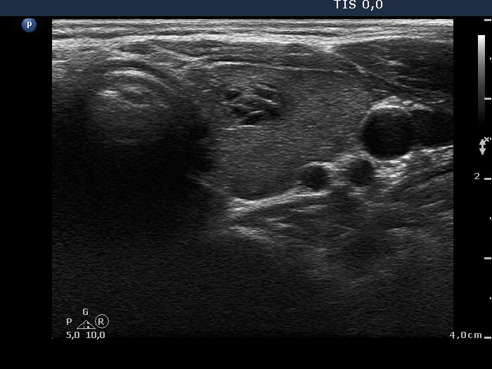
Benign hyperplastic nodule (histological diagnosis) - case 1662 |
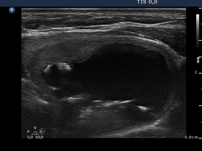
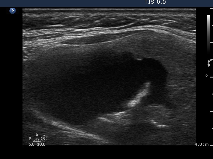
A cystically dilated macrofolliculus in a Graves' patient (cytological diagnosis) - case 969
|
 |
 |
 |
 |
The hyperechogenic lines and granules occur both within the parenchyma and dorsal to small cystic areas. The former correspond to connective tissue, the latter do to posterior back wall enhancement.
|
The hyperechogenic lines and granules are found almost exclusively dorsal to tiny cystic areas; therefore these belong to figures caused by posterior back wall enhancement.
|
| |
|
Benign colloid goiter (cytology) - case 15 |
|
 |
 |
 |
|
The lesion presents numerous hyperechogenic figures, including granules and lines. The figures are not related to ventral cystic areas, therefore these are presentations of connective tissue.
|
The presentation here is very similar to the case presented in left case. However, here the echogenic figures are in part dorsal to tiny cystic areas, therefore back wall figures should be considered first.
|
| |
|
| |
|
|
|
