
|
|
Benign nodular hyperplasia - Case 47.
|
|


Clinical presentation: a 19-year-old woman detected a nodule in her thyroid 2 weeks earlier.
Palpation: a firm solitary nodule in the right lobe.
Functional state: euthyroidism.
Scintigraphy indicated a "cold" nodule in the right thyroid.
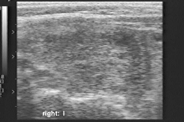
Ultrasonography: a moderately hypoechogenic nodule with lobulated margins occupied almost the entire right thyroid.
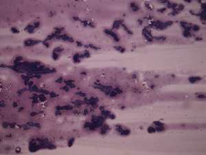
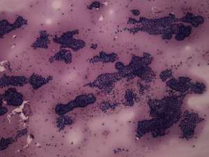
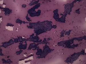
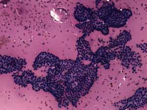
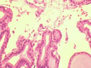
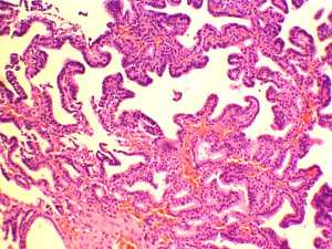
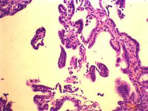
Cytological report suspicion of papillary carcinoma.
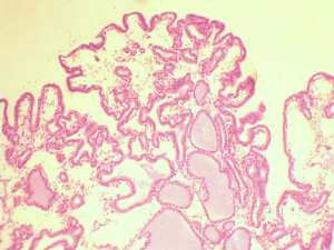
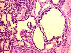
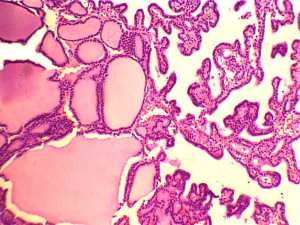
Histopathology: a normofollicular adenoma.
Comments.
-
This is a clear misinterpretation of the cytological pattern. The failure is explained first of all by the lack of enough cytological experience at the time of diagnosis. (We examined this patient in 1994 and this was our 1,307th thyroid FNAC. Now, in 2016 we are over 43,000 thyroid FNAC-s.) Moreover, this was a turning point in our cytological practice. We never again stained our smears with hematoxylin-eosin after the result of histopathology because of the weakness of this staining method to recognize nuclear inclusions and grooves.
-
The sonographic images are naturally out of date. Nevertheless, the lobulated margins of the nodule are remarkable.