|
|
|
 |
 |
|
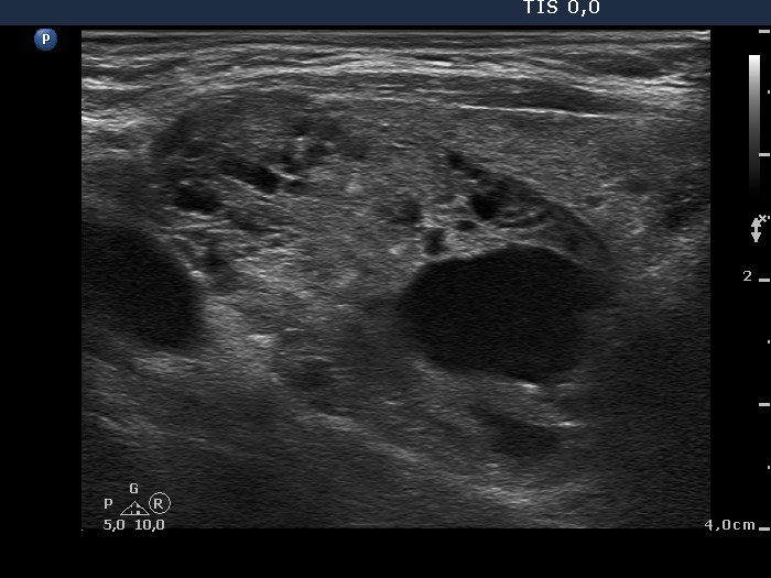
Note the presence of
microcalcifications at the edge of solid part.
|
Various hyperechogenic granules are
within the nodule.
|
|
|
|
|
|
|
|
 |
 |
 |
 |
|
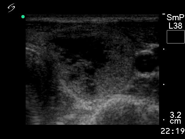
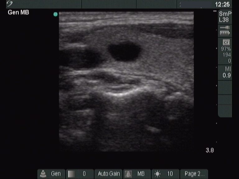
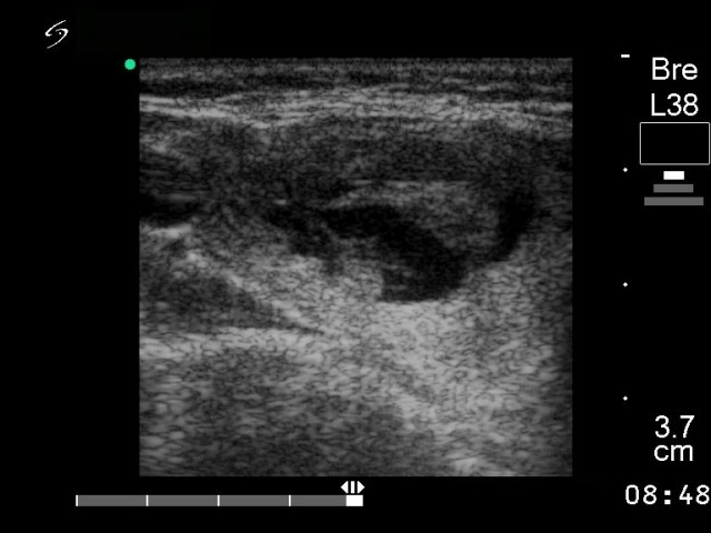
After evacuation of the seemingly
simple cyst a small suspicious solid part appeared with
microcalcifications.
|
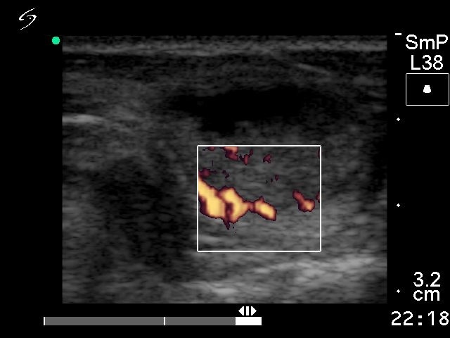
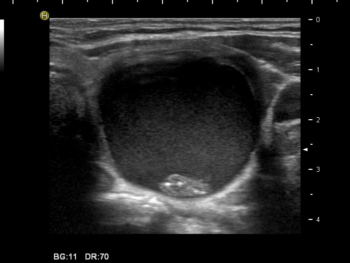
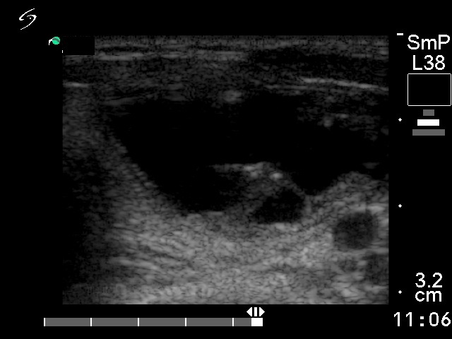
The dorsal solid part was unchanged
after evacuation of the cyst. It is also suspicious because of the
hyperechogenic granules.
|
| |
|
|
|
Benign hyperplastic nodules - Case 11 |
 |
 |
 |
 |
|
The shape of the solid part has no
relevance. Compare the appearance of hyperechogenic granules in these
cases. The hyperechogenic granules are more bright in the case of
papillary carcinoma compared to benign nodules. Nevertheless, this
difference does not have enough practical value.
|
| |
|
|
|
Benign hyperplastic
nodules - Case 34 |
 |
 |
 |
 |
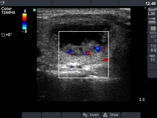
| The presence of microcalcifications and
the type 3 vascular pattern increased the possibility of being this
nodule malignant. |
The lack of vascularization on Doppler
mode has no relevance in the differential diagnostic of nodular
goiters. |
| |
|
|
|
Benign hyperplastic nodules - Case 13 |
 |
 |
 |
 |
| The only suspicious sign in the maliginant case is the macrolobulation of the solid part. Both cases belong to the peripheral-type of cysts. The vascularization is just the opposite of what we expect: the malignant nodule presents a type 2 perinodular while the benign lesion does a type 3 intranodular vascular pattern. |
| |
|
Taking all into account, the sonography
is not able to differentiate between benign and malignant cystic
nodules in general. However, the combination of lack of
microcalcifications and the type 3 vascular pattern, and the presence
of a halo significantly decreases the risk of malignancy. Another
important consideration: the echogenicity of the solid part is
optically influenced by the presence of the cystic fluid. It means that
in contrast with solid nodules, the echogenicity of the solid part in
mixed nodule has no relevance. Malignancy can be found with equal
probability in a mixed nodule with echonormal and hypoechogenic solid
parts.
|