Benign hyperplastic nodules - Case
24 |
 |
 |
|
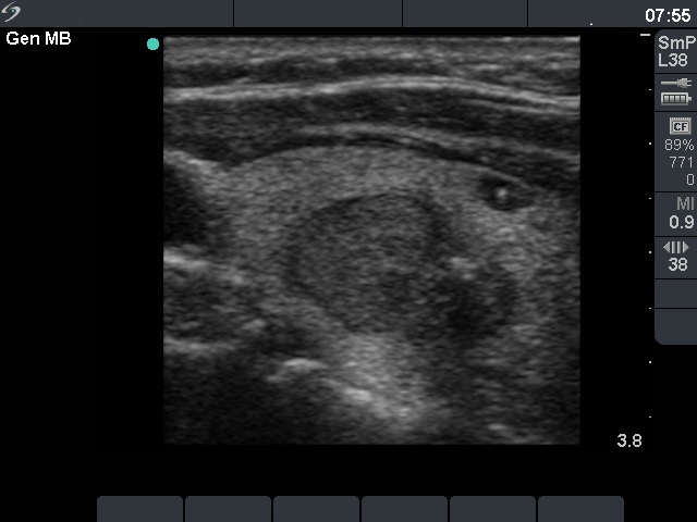
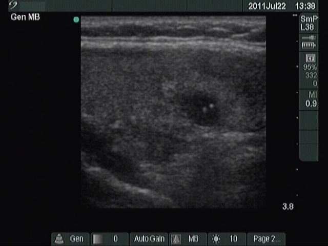
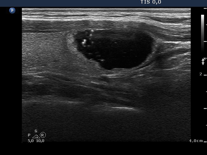
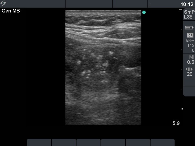
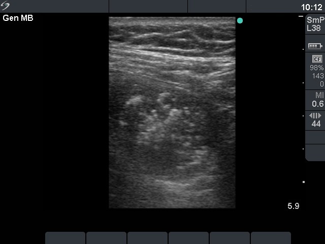
The images demonstrate four types of intranodular hyperechogenic
figures: microcalcifications marked with red, macrocalcifications with
the characteristic dorsal acoustic shadow marked with blue, comet tail
artifacts marked with yellow and patchy hyperechogenic figure marked
with green. The latter is a relatively specific feature of amyloid
deposit, but may be observed in other thyroid lesions, as well.
|
| |
|
|
|
 |
 |
|
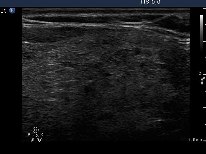
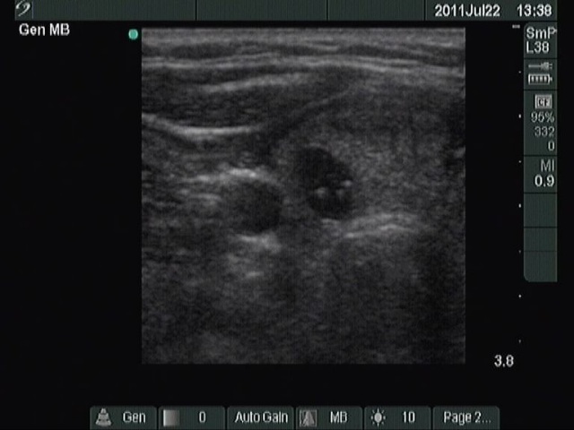
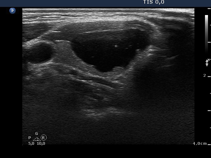
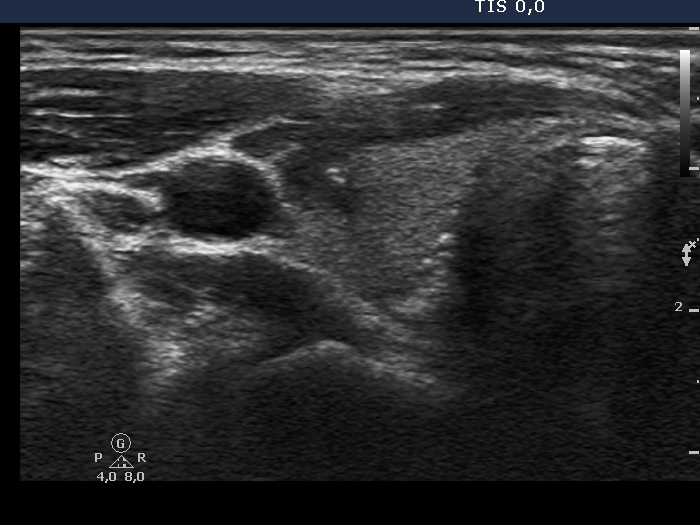
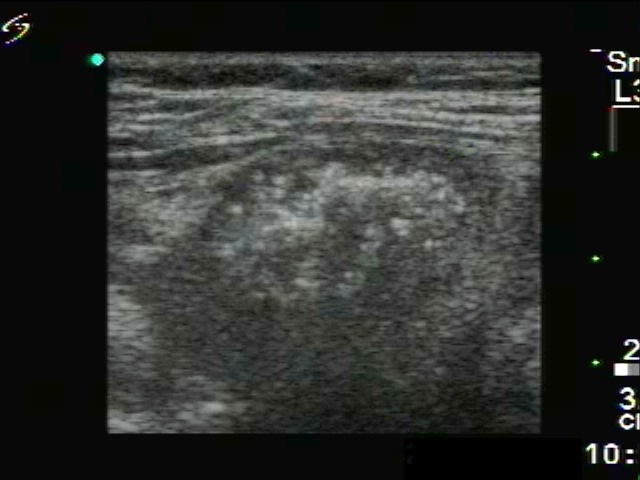
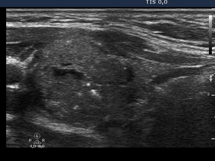
Three distinct pathological entities were diagnosed on histology. The
largest nodule presenting halo sign proved to be an oxyphilic
adenoma, the smaller lesion next to the former containing
microcalcifications marked with red arrow was a papillary cancer, while
the smallest lesion with a comet-tail artifact marked with yellow was a
benign hyperplastic nodule. Note the similarity of the different
hyperechogenic granules. This case illustrates why is it so difficult
to differentiate these granules in a great proportion of cases.
|
| |
|
Benign hyperplastic nodules - Case
1 |
 |
 |
|
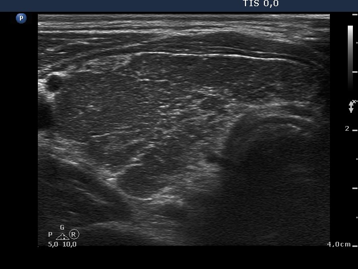
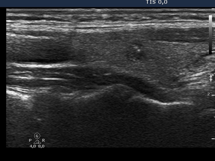
Fibrosis is presented as granules or lines depending on the angle
between the fibrotic bundle and the ultrasound wave. If both
hyperechogenic granules and lines are found, it is fibrosis with great
probability. If we are not aware of this possibility, we may
misinterpret a hyperechogenic granule as a microcalcification.
|
| |
|
|
|
 |
 |
 |
 |
|
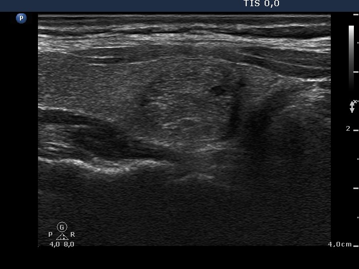
Graves' disease frequently presents extensive proliferation of connective tissue.
Fibrotic bundles draw the subunits of the lobe visible. In contrast with the extranodular part, the lesion in the left lobe does not present fibrosis.
|
| |
|
Benign hyperplastic nodules - Case
12 |
|
 |
 |
 |
 |
|
A nodule with extensive fibrosis is
presented.
|
A papillary cancer with numerous
microcalcifications is demonstrated
|
|
The two cases
differ in the ratio of hyperechogenic granules to lines. In the case of
fibrosis the ratio is close the 1, while in the case of
microcalcification granules predominate over lines. In the event of the
former the presentation of the hyperechogenic figures depends on the
angle between the ultrasound and the linear fibrotic bundle and
therefore only the chance influences the appearance of the fibrosis.
Moreover, microcalcifications itself are round figures.
|
| |
|
Benign hyperplastic nodules - Case
42 |
Benign hyperplastic nodules - Case
46 |
 |
 |
 |
 |
|
The hyperechogenic granules demonstrated in the images are the
so-called comet-tail artifacts. They probably represent areas rich in
colloid and are found characteristically in benign hyperplastic nodules.
|
| |
|
Benign hyperplastic nodules - Case
15 |
|
  |
  |
The presentation hyperechogenic granules is very similar in both of the above cases. If we had known the final diagnosis, it would have been very simple to interpret
the hyperechogenic granules in the left, benign lesion as
comet tail artifacts, while those in the right, malignant case as
microcalcifications. To differentiate comet-tail artifacts from
microcalcifications in reality is much easier than analyzing images. |
| |
|
Ethanol sclerotherapies - Case 2 |
|
 |
 |
 |
 |
|
Small bright hyperechogenic granules are presented. These may be
microcalcifications. The correct interpretation of small hyperechogenic
granulations is more successful in real-time than in images.
|
| |
|
Benign hyperplastic nodules - Case
52 |
Benign hyperplastic nodules - Case
8 of a new approach |
 |
 |
 |
 |
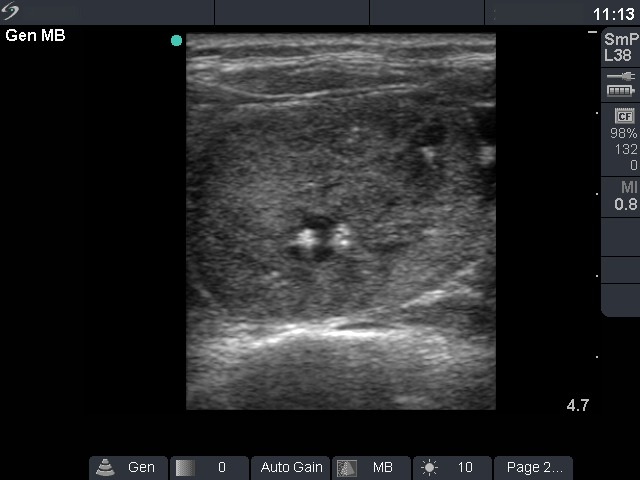
| The nodules display coarse calcifications
with the pathognomic dorsal acoustic shadow. In the right case, the
presence of acoustic shadow is unequivocal in color mode. |
| |
|
Benign hyperplastic nodules - Case
18 |
|
  |
  |
|
The so-called eggshell calcification is demonstrated in these two
cases. This sonographic property increases the possibility of carcinoma
in statistical manner. Nevertheless, the practical relevance of this
sign is not so great.
|
| |
|
Benign hyperplastic nodules -
Case 21 |
|
  |
  |
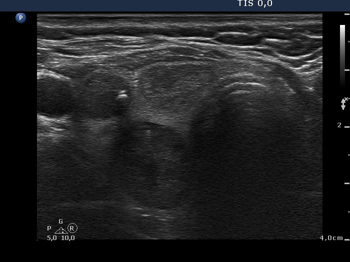
Larger, cotton-like hyperechogenic structures are demonstrated. These
may be the sonographic presentation of amyloid deposit and therefore
the sign of medullary cancer. |
| |
|
|
|
 |
 |
 |
 |
Various forms of amyloid deposit are presented. In the left case smaller
while in the right case larger hyperechogenic patches are demonstrated.
|
| |
Granulation around surgical thread - Rare forms of
theyroiditis - Case 2 |
  |
  |
|
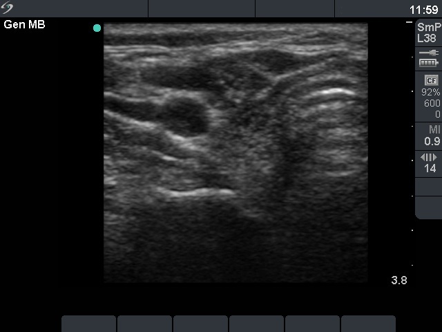
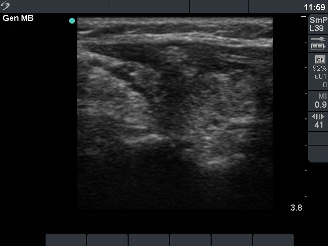
The presentation of granulation around surgical thread is very similar to
that of an amyloid deposit: there are patchy and granular hyperechogenic figures
in a hypoechogenic background. These lesions are always avascular and
the irregular shape of the whole lesion are of help. The patient
history is the clue of the diagnosis. These granulations are very hard
and painless.
|
| |
Granulation around surgical thread - Rare forms of theyroiditis - Case 4 |
 |
 |
|
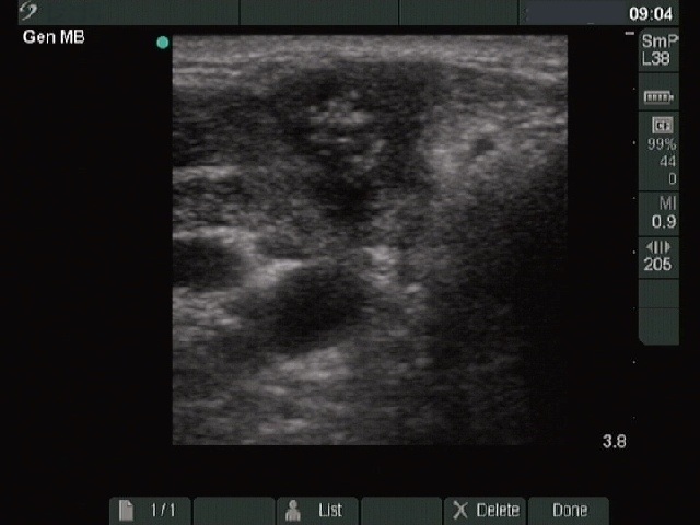
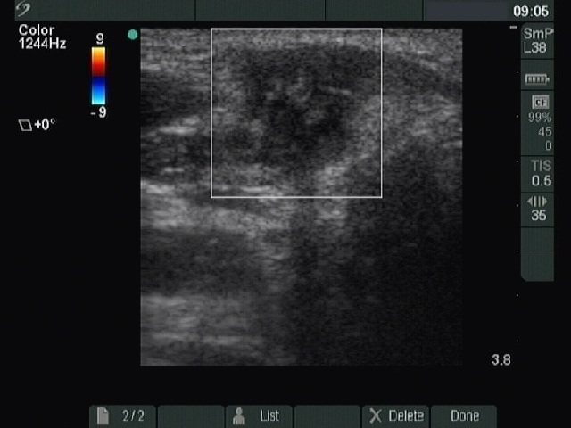
Note the irregular shape of the lesion and the lack of vascularization
on Doppler mode. The presentation of granulation around surgical thread
is similar to that of an amyloid deposit: relatively larger patches with small hyperechogenic granules can be found
in a hypoechogenic background.
|

